Core Vaccination Program and Infectious Disease Control for Horses
Core Vaccination Program and Infectious Disease Control for Horses
Authors: Fernanda Camargo and Kristen Harvey, Department of Animal Sciences
Introduction
Programs for the control of infectious diseases are important components of good management practices directed toward maximizing the health, productivity, and performance of horses. Infectious diseases occur when horses are exposed to a sufficient quantity of an infectious agent that overcomes the horses' previously acquired resistance.
Some of the goals of disease control programs include the reduction of factors that may decrease resistance or increase susceptibility of disease in the horse. Such factors include stress, overcrowding, parasitism, poor nutrition, inadequate sanitation, contaminated water source/supply, concurrent disease, stringent exercise program, no turn-out time, transportation, inclement weather, weaning, disruption of established social groups, and movement of people, vehicles, and/or equipment on and off facilities during infectious disease outbreaks. Consistent utilization of good management programs will lower the incidence and/or severity of infectious diseases in any particular herd.
A well-delineated vaccination program can also enhance resistance to those diseases; however, vaccines can protect horses from some, but not all, infectious diseases. A horse or groups of horses that are well fed and well managed are more likely to maintain good health.
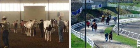
The occurrence of infectious diseases tends to increase at sales or boarding facilities, breeding farms, barns of performance and show horses, fairgrounds, or racetracks. Such situations are ideal for introducing and transmitting infectious diseases, particularly infections of the respiratory tract.
In farms where there is a resident herd and an itinerant herd, the management of the two distinct populations needs to be taken into account. The introduction of horses from various backgrounds and origins and the commingling of horses of different ages, career/use, and traveling status create special problems and need to be specifically addressed in the practice of disease control.
Management recommendations are based on the practices of (1) segregation of horses on the premises into small groups, (2) the maintenance of each horse group as an isolated unit, and (3) stress reduction. Horses should be segregated into groups that are similar in age, gestation status, use, and frequency of removal of individual animals. Restrictions should be placed on movement of horses into and out of each established group, and contact with transient horses in particular should be avoided. The greatest danger lies in the introduction of new horses into established groups, especially those with recent opportunities for exposure to large, intermingled gathering of horses from diverse sources (sales, shows, boarding stables, racetracks, competitions, training centers, etc.).
Ideally, horses should be appropriately vaccinated at least one month prior to entering or leaving such a facility in order to produce adequate antibodies before the anticipated exposure. Ideally, before a new horse is introduced into a closed group, it should be isolated for 21 days. In addition, a horse temporarily removed from a group for prolonged transport or contact with other horses (e.g., breeding, showing, training, veterinary care, sales, etc.) should also undergo a 21-day isolation period and evaluation for signs of infection before being returned to its resident group.
In cases of respiratory and digestive system diseases, infection often spreads rapidly, resulting in significant economic impact. Immediate intervention with infectious disease control measures is required for successful containment of the infection. Implementing the practices of isolation, quarantine, and disinfection is imperative for managing an outbreak. Clinically ill horses should be removed from their resident group and kept in physical isolation away from the remainder of the horse population. A zone of empty space sufficient to serve as a barrier should surround the isolation area. Human and horse traffic through the isolation area should be restricted. Personnel caring for horses placed in isolation should wear protective outer clothing, disposable latex gloves, and footwear that can be immersed in a disinfectant footbath. All of the outer protective clothes should be removed upon leaving the isolation area. Ideally, staff handling horses in isolation would be assigned to that group only. Remember to thoroughly clean and disinfect the area used as isolation facilities after resolution of the disease.
Vaccination
Vaccination is also an important part of infectious disease control.
The American Association of Equine Practitioners (AAEP) and the American Veterinary Medical Association (AVMA) divide their vaccination program into two categories: core and risk-based vaccination guidelines. The AVMA defines core vaccinations as those “that protect from diseases that are endemic to a region, those with potential public health significance, required by law, virulent/ highly infectious, and/or those posing a risk of severe disease.” It is recommended that all horses receive the vaccinations included in this group. See tables below for specific age or gestation status before vaccinating your horses. Diseases included in the core list are tetanus, rabies, eastern and western equine encephalomyelitis, and West Nile virus. Diseases included in the risk-based list vary regionally, from population to population within an area, or even between individual horses within a population. Diseases included in the risk-based list are anthrax, botulism, equine herpesvirus (rhinopneumonitis), equine viral arteritis, equine influenza, Potomac horse fever, rotaviral diarrhea, and strangles. It is important to consult your veterinarian to develop a vaccination protocol for your horse population. This publication will focus on the core vaccination guidelines.
Core Vaccination Guidelines
Tetanus
Tetanus is caused by the toxins of the anaerobic bacterium Clostridium tetani. C. tetani is a ubiquitous soil inhabitant and can be isolated from feces of horses and other domestic animals. Among animals, the horse is considered the most sensitive to tetanus toxin. Tetanus is an infectious disease but not a contagious disease, and, as such, it cannot spread from animal to animal or from animal to humans.
The most common route of infection is puncture wounds contaminated with manure and soil that contain C. tetani spores. However, causative wounds may not be found in up to 30% of tetanus cases.
C. tetani needs an anaerobic environment to multiply and produce toxins, and that is why deep penetrating (or puncture) wounds produce the most favorable circumstances for the organism to flourish and produce the toxins that will lead to the disease. However, tetanus can develop after minimal superficial damage, provided an anaerobic state develops in the affected area.
Tetanus toxins act in a way that all muscles in the body contract simultaneously when stimulated, in contrast to the normal state where contraction of one muscle is accompanied by relaxation in its counterpart. This shows up clinically as muscle rigidity and muscle spasms. The force of these contractions may cause rhabdomyolysis (the breaking down of muscles), intramuscular hemorrhage, fracture of long bones, etc. The spasmodic (called tonic-clonic) convulsions are very strenuous to the affected animal, and the resulting hemodynamic instability is an important cause of death.
Clinical signs of tetanus include rigidity, stiff and stilted gait, elevated tail, persistent protrusion of the third eyelid, flared nostrils, excessive reaction to sound and light, profuse sweating, and a “sawhorse” stance with legs in a rigid extended position. As the disease progresses, the horse develops “lockjaw” (sardonic smile, or risus sardonicus) where the jaw muscles are rigidly contracted. The horse will be unable to drink or eat. If untreated, the involvement of the respiratory muscles and the generalized musculoskeletal injuries will lead to death in more than 80% of cases.
The diagnosis of tetanus is based on clinical signs. Treatment involves the provision of a safe and quiet environment, elimination of unbound toxin (by administration of anti-toxin), sedation and muscle relaxation, and general support (intravenous fluids, antibiotics, etc). In some cases, euthanasia needs to be considered.
Tetanus has long been preventable by vaccination with inexpensive tetanus toxoid. These vaccines induce long-lasting immunity. Primary vaccination involves the administration by intramuscular injection of two initial doses (1 month apart) and a yearly booster. For unvaccinated horses that sustain an injury, an optional approach is to administer the tetanus antitoxin and the vaccine at the same time (in different muscles). The antitoxin will provide immediate protection that will last for up to 4 weeks. The second dose of vaccine should be administered 4 weeks later to complete the primary series. Thereafter, the annual boosters are imperative for protection.
Rabies
Rabies virus causes acute encephalitis in all warm-blooded hosts including humans, and the outcome is almost always fatal. Although all species of mammals are susceptible to rabies virus infection, only a few species are important as reservoirs for the disease. Transmission of rabies usually occurs through the saliva from an infected animal's bite. The wildlife species that serve as the natural reservoir for infection vary between different parts of North America and include raccoons, skunks, foxes, badgers, and bats. Bites on horses are more frequently on the muzzle, face, and lower legs. The virus then reaches the spinal cord and brain through the nerves and causes rapidly progressive and fatal encephalitis. In 2006, there were a total of 53 cases of rabies in horses in the United States, reported by the Centers for Disease Control. A total of 6,943 cases
of rabies in animals were reported for 2006.
The clinical signs range from strange behavior, lameness, neurological deficits, self-mutilation, fear, agressiveness, and depression, etc. Clinical signs are progressive from onset until death, which
generally happens by day 10.
There is no definitive antemortem (before death) test for rabies in horses. Clinical signs are suggestive but non-diagnostic. The diagnosis is made by postmortem evaluation of the brain of
the suspected animal.
There is no effective treatment for horses (and other animals, vaccinated or unvaccinated) with clinical rabies. Horses w it h cl i n ica l sig n s of rabies should be isolated to prevent human exposure to the virus.
One of the most important things about rabies is that it is considered a zoonotic disease and, as such, is transmissible from animals to humans. Animals with suspected rabies should only
be handled by trained individuals who have had the appropriate rabies vaccination.
All horses, regardless of breed, gender, or use, should be annually vaccinated against rabies. The unvaccinated horse should receive a primary set of three doses and annual boosters. Vaccinated
horses and broodmares should receive annual boosters.

Horses in poor body condition have a higher chance of acquiring an infectious disease.
|

Good management is imperative in the control of infectious diseases. Good management is imperative in the control of infectious diseases. |
Eastern and Western Equine Encephalomyelitis (Sleeping Sickness)
Sleeping sickness is a lay term used to describe a neurological disease caused by viruses of the Alphavirus genus. These viruses, causing agents of the Eastern and Western and Venezuelan equine encephalomyelitides (EEE, WEE, VEE), are arboviruses, meaning that they are transmitted by bloodsucking insects, such as mosquitoes. EEE and WEE receive their names because of the geographical location where they were more frequently associated with outbreaks in the United States. VEE causes outbreaks of encephalitis in horses in Central America, South
America, Mexico, and occasionally in the southern part of the United States.
Although widespread vaccination has reduced the size and number of outbreaks of EEE and WEE in horses, the impact of these diseases is still significant because of the rapid and severe nature of clinical signs and high mortality rate in affected horses. EEE in the United States has been detected in all states east of the Mississippi River and in some western states. In total, several hundred equine cases and about 10 human cases are reported each year.
The life cycle of these viruses involves the transmission between birds, rodents. and mosquitoes. Humans are also susceptible to these diseases when the virus is transmitted to them by infected mosquitoes. Ticks and many species of mosquitoes are capable of virus transmission. Of these encephalitides, WEE has the lowest mor t a l it y i n hors es (approximately 50%). EEE is the most virulent for horses, with mortality approaching 90%.
VEE is a reportable foreign animal disease. As such, vaccination against VEE is not recommended in most parts of the United States, including Kentucky.
The clinical signs for these diseases are those of central nervous system (CNS) involvement, including ataxia, paralysis, irregular gaits, grinding of teeth, incoordination, circling, staggering, head pressing, partial or total blindness, head hanging low with drooping ears and lips, and ultimately stupor. The profound depression associated with these infections give rise to the common name of "sleeping sickness." The course of disease varies between 2 and 14 days. Nearly all horses with EEE will die, whereas horses with WEE have a higher chance of survival.
The diagnosis is made by analyzing the cerebrospinal fluid (CSF) of the affected horse or isolating the virus from the brain of the dead horse. Blood samples are not useful for virus recovery because usually no circulating virus is present when signs of encephalitis appear. It is not possible to diagnose EEE or WEE by the clinical signs alone, and currently there are no reliable antemortem specific diagnostic tests available.
West Nile Virus
The family Flaviviridae, to which the West Nile virus (WNV) belongs, has the distinction of containing some of the most important human pathogens in the world. Some of the viruses in this
family include the causing agents for yellow fever, dengue, Japanese encephalitis, tick-borne encephalitis, louping ill, etc.
| Broodmares | Adult Horses (>1 year of age) | Coments | ||
| Vaccinated: | Unvaccinated:1 | Vaccintated: | Unvaccinated:1 | |
| Tetanus | Tetanus | Tetanus | ||
| Annual, 4-6 weeks prepartum |
|
Annual |
|
Booster at time of penetrating injury or prior to surgery if last dose was administered over 6 months previously. |
| EEE/WEE | EEE/WEE | EEE/WEE | ||
| Annual, 4-6 weeks prepartum |
|
Annual, spring, prior to onset of vector season |
|
Consider 6-month revaccination interval for:
|
| WNV | WNV | WNV | ||
| Annual, 4-6 weeks prepartum |
It is preferable to vaccinate unvaccinated mares when open. In areas of high risk, initiate primary series as described for unvaccinated adult horses. |
Annual, spring, prior to onset of vector season | Inactivated vaccine:
Recombinant canary pox vaccine:
Flavivirus chimera vaccine:
|
When using the inactivated or the recombinant product, consider 6-month revaccination interval for:
|
| Rabies | Rabies | Rabies | ||
| Annual, 4-6 weeks prepartum OR prior to breeding |
Annual, 4-6 weeks prepartum OR prior to breeding |
Annual | Single dose, annual revaccination |
Due to the relatively long duration of immunity, this vaccine may be given post-foaling but prior to breeding and thus reduce the number of vaccines given to a mare prepartum. |
1 Unvaccinated or lacking a vaccination history.
West Nile virus is the leading cause of arbovirus encephalitis in horses and humans in the United States. Since 1999, more than 25,000 cases of W N V encephalitis have been reported in U.S. horses. The occurrence of more than 3,000 human cases in 2007 indicates widespread viral activity in the environment.
This virus has been identified in all of the continental United States, most of Canada, and Mexico. Several Central and South American countries have also identified WNV within their borders.
The virus is transmitted from avian reservoir hosts by mosquitoes (and infrequently by other bloodsucking insects) to horses, humans, and a number of other mammals. West Nile virus is transmitted by many different mosquito species, and this varies geographically. Horses and
humans are considered to be dead-end hosts for WNV, which means that the virus is not directly contagious from horse to horse or horse to human. Indirect transmission via mosquitoes from
infected horses is unlikely as these horses do not circulate a significant amount of virus in their blood.
The mortality rate in affected horses is 30 to 35%. Many horses will improve within 3 to 7 days of displaying clinical signs. If the horse demonstrates significant improvement, full recovery can be
expected within 1 to 6 months. Some horses will persistently present residual weakness, ataxia, gait, and behavioral abnormalities, etc.
| Foals and Weanlings (< 12 months of age) | Comments | |
| Of mares vaccinated in the prepartum period:1 |
Of unvaccinated mares: | |
| Tetanus | Tetanus | Tetanus |
3-dose series:
|
3-dose series: • 1st dose at 1-4 months of age • 2nd dose 4 weeks after the 1st dose • 3rd dose 4 weeks after second dose |
|
| EEE/WEE | EEE/WEE | EEE/WEE |
|
3-dose series:
*Foals in the Southeastern U.S.: The primary |
3-dose series:
*Foals in the Southeastern U.S.: The primary |
Note: Primary vaccination series scheduling may be amended with vaccinations administered earlier to younger foals that are A foal born during the vector season may |
| WNV | WNV | WNV |
| Inactivated vaccine:* 3-dose series:
*Foals in the Southeastern U.S.: Due to early seasonal vector presence, the primary |
Inactivated vaccine:* 3-dose series:
* Foals in the Southeastern U.S.: Due to early seasonal vector presence, the primary vaccination series should be initiated at 3 months of age. |
ote: Primary vaccination series scheduling may be amended with vaccinations administered to younger foals that are at increased risk of exposure due to the presence of vectors. |
| Recombinant canary pox vaccine: 3-dose series:
|
Recombinant canary pox vaccine: 3-dose series:
|
A foal born during the vector season may warrant initiation of the primary vaccination series at an earlier age than a foal born prior to the vector season. |
| Flavivirus chimera vaccine: 2-dose series:
|
Flavivirus chimera vaccine: 2-dose series:
|
There are no data for the use of the recombinant or chimera product in foals < 5 months of age. If either product is administered to foals at < 5 months of age, the recommended primary schedule should still be completed. |
| Rabies | Rabies | Rabies |
3-dose series:
|
3-dose series:
|
|
The clinical signs of WNV infection are that of CNS involvement, which includes change of personality, hyperexcitability, depression, aggressiveness, etc. Some horses can show signs of sudden sleep-like activity resembling narcolepsy. Muscle twitching is also observed and can involve all four limbs and trunk,which will affect normal activities such as walking, eating, and interaction with handlers. Some horses present a short, slow, stilted gait which sometimes is confused with lameness. They can also show signs of ataxia and incoordination. They may collapse and be unable to get up, requiring aggressive supportive care.
It is impossible to diagnose WNV from clinical signs alone, as many other diseases that affect the CNS present the same clinical signs. There are some laboratory tests that can be run in an attempt to diagnose WNV, including complete blood work, serum biochemistry, CSF analysis, and other tests. There are also postmortem methods for confirming WNV infection. There are three types of West Nile virus vaccines, so you need to discuss with your veterinarian about which one
is the most appropriate for your herd. Other considerations on arboviruses include the use of mosquito repellent and elimination of mosquito breeding sites (water in flowerpots, pet bowls, buckets, barrels, discarded tires, and other items that could collect water).
References
American Association of Equine Practitioners (www.aaep.org).
Centers for Disease Control and Prevention, Division of Vector-Borne Infectious Diseases, Eastern Equine Encephalitis (http://www.cdc.gov/ncidod/dvbid/Arbor/eeefact.htm).
Centers for Disease Control and Prevention, Division of Vector-Borne Infectious Diseases, West Nile Virus (https://www.cdc.gov/ncidod/dvbid/westnile/).
Centers for Disease Control and Prevention, Rabies (http://www.cdc.gov/RABIES/).
Gibbs, E.P.J., Long, M.T. Equine Alphaviruses. In: Equine Infectious Diseases. Editors: Sellon D.C. and Long, M.T. Saunders-Elsevier, Saint Louis, Mo., pp. 191-197, 2007.
Long, M.T. Flavivirus Infections. In: Equine Infectious Diseases. Editors:Sellon, D.C. and Long, M.T. Saunders-Elsevier, Saint Louis, Mo., pp. 198-206, 2007.
MacKay, R.J. Tetanus. In: Equine Infectious Diseases. Editors: Sellon, D.C. and Long, M.T. Saunders-Elsevier, Saint Louis, Mo., pp. 376-380, 2007.
United States Department of Agriculture, Eastern Equine Encephalomyelitis (http://www.aphis.usda.gov/lpa/pubs/fsheet_faq_notice/fs_aheasterne.html).
Wilkins, P.A., Del Piero, F. Rabies. In: Equine Infectious Diseases. Editors: Sellon, D.C. and Long, M.T. Saunders- Elsevier, Saint Louis, Mo., pp. 185-1191, 2007.
Wilson, W.D., Spensley, M.S. Preventive Medicine Programs. In: Current Therapy in Equine Medicine 3. Editor: Robison, N.E. W.B. Saunders, Philadelphia, Pa., pp. 35-50, 1992.
