Equine Metabolic Syndrome: Is My Horse Just Fat or Is He Sick?
Equine Metabolic Syndrome: Is My Horse Just Fat or Is He Sick?
Authors: Amanda Adams, Veterinary Sciences, and Fernanda Camargo, Animal and Food Sciences
How Do We Define Equine Metabolic Syndrome?
Equine Metabolic Syndrome (EMS) is an endocrine disorder that affects equids (horses, ponies, and donkeys) in three defining ways:
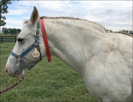
- They are obese and/or have localized fat deposits (Figure 1).
- They are in an insulin resistance (IR) state, now referred to as insulin dysregulation (ID).
- They are predisposed to developing laminitis.
Unfortunately, we have limited understanding of the pathophysiology or cause of EMS due to its complexity and the involvement of both metabolic and inflammatory pathways, whose interactions are poorly understood.
How Does It Happen?
EMS often results from genetic predisposition combined with environmental factors, including overfeeding and lack of exercise, especially in the “easy keeper” horse. These horses can become obese and usually exhibit increased regional adiposity. Chronic obesity can develop in horses when they reach maturity and may continue for more than 20 years. Obesity in horses is most often defined using the Henneke Body Condition Scoring system, which rates horses from 1 (poor) to 9 (obese). More precise measurements of body fat mass can also be obtained by measuring rump fat thickness or with ultrasound. Obesity is associated with increased adiposity, which often times contributes to insulin resistance. How? Adipose tissue is an endocrine organ made up of fat cells called adipocytes. Adipocytes are capable of producing hormones and inflammatory cytokines, both of which can contribute to insulin resistance.
What is Insulin Resistance and what’s the Big Deal about It?
Equids affected by EMS have a problem with the release and function of a key hormone in the body called insulin. Normally after a horse eats a meal, these nutrients are broken down into glucose which stimulates the pancreas to produce insulin. The insulin will then bind to insulin receptors on cells in metabolically active tissues (skeletal muscle, adipose, and liver), which triggers a series of intracellular events that ultimately leads to rapid glucose uptake into these tissues. Basically insulin helps feed these metabolically active tissues so that they stay healthy, in turn keeping the horse healthy and active. Now, in an equid affected by insulin resistance or decreased insulin sensitivity, this process we just described is not happening normally and can simply be defined as a failure of insulin to stimulate tissue glucose uptake when nutrients are abundant after feeding. Thus, these EMS horses live in a state of hyperinsulinemia (higher than normal levels of insulin circulating in blood) because the pancreas is producing more insulin than needed in order to overcompensate for the failure of tissues to respond to insulin. This hyperinsulinemia over time decreases the sensitivity of tissues to insulin and leads to lower tissue glucose uptake. This tissue insulin resistance causes an increase in insulin secretion by the pancreas, especially after a meal, and thus further perpetuates the cycle. IR in the horse is similar to metabolic abnormalities that accompany type II diabetes in humans. IR can also be seen in horses suffering from PPID (Pituitary Pars Intermedia Dysfunction), formerly known as Equine Cushing’s Syndrome.
The problem or why insulin resistance is so concerning is that it is a contributing factor to EMS horses developing laminitis. Laminitis is a devastating and debilitating condition of the hoof, often times resulting in euthanasia. The tissues within the hoof (laminae) that anchor the coffin bone to the hoof wall become weakened by hormones, abnormal metabolic state, and inflammation. Because these laminae tissues become weak, this allows the bone to rotate in the hoof capsule causing chronic lameness or to a degree which warrants euthanasia. What we often times do not realize is the fact that EMS horses can have mild laminitis events causing cumulative damage over time, leading to more severe laminitis. Thus, it is critical to detect EMS early.
Is Every Horse at Risk?
While horses of any breed may develop EMS, ponies, donkeys, and horse breeds that are considered easy keepers have higher propensity to develop EMS and become insulin resistant.
What Does EMS Look Like?
EMS horses are those that become obese more easily than others. They develop fat deposits on the neck, termed cresty neck (Figure 2); around the tail head (Figure 3) and loin; and many times around the utter and prepuce. These regional adiposity areas can occur even if the rest of the body looks less obese or even normal. Some horses may have a regional fat deposit while others appear fat in general. Therefore, obesity alone as determined by BCS is not a definitive diagnosis of EMS. Indeed, some obese horses have normal levels of resting insulin while horses of a normal weight with some regional adipose deposits may be insulin resistant. Further, because EMS and PPID share similarities, in that both diseases can cause regional adipose tissue deposits, laminitis and IR, it is highly important to differentiate their diagnosis as treatment is not the same. To complicate matters even further, sometimes the same horse will test positive for both EMS and PPID. Thus, it is critical to perform appropriate endocrine diagnostic testing to determine and not guess the endocrine status of the horse.

How Do We Diagnose EMS?
Diagnosis is based on history, clinical signs, and blood tests.
The best measurement of insulin dysregulation is performed by determining fasted, resting insulin levels and insulin responses to a dynamic test, called the oral sugar test. We can measure insulin in the fasted horse, which involves giving the horse nothing to eat following a flake of hay at 10 p.m. and collecting blood first thing the following morning. A minimum of 6-8 hours fasting is recommended. Morning insulin and glucose levels are measured. Hyperinsulinemia is indicative of IR. Although this test is easy, it is less sensitive than the oral sugar test and may not identify early cases of EMS.
The oral sugar test involves drawing blood in the morning, followed by giving the horse a dose of Karo syrup (the dose is calculated based on body weight) via oral syringe, and collecting blood at 60 minutes and sometimes 90 minutes after the oral dose. Insulin and glucose are measured in the morning and following the oral dose at the 60 or 90 minute time points. High insulin levels at the 60 or 90 minutes sample collection will confirm the diagnosis. It is critical that your veterinarian works with appropriate endocrinology labs in order to ensure correct blood results.
Before having your vet perform any of these tests, it is important to know that insulin normally increases after feeding, as well as during stress (secondary to cortisol increase) and in response to pain (secondary to epinephrine increase). Therefore, insulin tests should be avoided if any of these conditions are present, such as during an acute laminitis episode.
How to Treat EMS?
The main goal of treatment is to encourage weight loss. Weight loss is generally accomplished by decreasing caloric intake and increasing caloric expenditure. This can be done by dietary changes and increases in exercise. However, some horses may also need to be treated medically to induce weight loss especially if they are unable to exercise or resistant to weight loss by diet. Your veterinarian will know the type of medication needed and dosage based on horse’s current weight and blood test results.
In general, EMS horses need to have a very restricted diet, with limited amounts of grass by either a dry lot condition or grazing muzzle, elimination of concentrates, sugary feeds and treats, and the addition of exercise (if the horse is currently sound and not having a recurrent bout of laminitis). A vitamin and mineral supplement, or ration balancers, can be added to the diet to ensure adequate nutrition if the horse is on a hay diet only. Once a horse loses weight, most can resume some pasture turnout as long as they wear a grazing muzzle. For tips on how to help your horse lose weight, see ASC-187: Help! My Horse Is Too Fat!
Research has shown that decreasing body fat not only improves insulin sensitivity but dampens inflammatory cytokine production, both components of EMS. There are other substances and alternative therapies that have been shown promising results in humans, but very little research has been conducted in the horse. Some nutritional supplementation has been shown in humans to have effects on insulin resistance. Effective supplements include different classes of polyphenols such as curcuminoids and resveratrol as well as prebiotics, probiotics, and certain fatty acids such as omega-3 fatty acids.
Take-home Message
EMS is a disease that can lead to devastating consequences such as euthanasia. It occurs more often in horses that are visibly easy keepers and overweight, but can also happen in horses that only have localized fat pads. The treatment includes weight loss and weight management. However, the effectiveness of any weight loss program depends on the willingness of the owner to not only implement the process but to continue these dietary changes over the life span of the horse. Work with your veterinarian and farrier to design a customized management plan tailored to your horse’s individual needs and to keep him sound with a good quality life.
