Wobbler Syndrome in Horses
Wobbler Syndrome in Horses
Authors: Fernanda C. Camargo, Animal and Food Sciences, and Jennifer Janes, Veterinary Science
Introduction
Wobbler syndrome, or cervical vertebral malformation (CVM), is a devastating disease that can affect a horse’s neurologic and musculoskeletal systems. It is a structural narrowing of the spinal canal due to a variety of vertebral malformations and leads to spinal cord compression. As a result, horses exhibit clinical signs of spasticity, ataxia, and lack of coordination.
Cervical vertebral instability and cervical static stenosis are the two distinct types of CVM. Cervical vertebral instability causes dynamic spinal cord compression—occurring when the neck is flexed. Horses with dynamic lesions are typically 4 to 18 months of age, and the location of the compression is typically C3-5 (between the cervical vertebrae 3 and 5).
Cervical static stenosis typically affects horses between 1 and 4 years of age and is characterized by narrowing of the cervical canal C5-7 (between the cervical vertebrae 5 and 7). The compression that results is static—occurring regardless of neck position.
These distinctions of dynamic versus static compression are made on the basis of a myelogram, a diagnostic technique that is discussed later in this publication. It is important to note the distinction between these two classifications is not always clear. Horses have been reported to have a narrowed canal (associated with cervical static stenosis) extending from C3 to C7 (including vertebrae generally associated with both cervical vertebral instability and cervical static stenosis).
This disease has been reported in a number of breeds, with Thoroughbreds, Quarter Horses, and Warmbloods being those most commonly affected. Th Miniature Horse is the only breed that has escaped diagnosis of wobbler syndrome.
Typically, wobbler syndrome is thought of as a developmental disease of young horses and is even included in the classification of developmental orthopedic diseases by some authors. Researchers have looked at this disease in horses over the age of 4 and concluded that it should be kept as a differential diagnosis in older horses as well.
Causes
The exact cause of wobbler syndrome in horses is unknown. It is believed that the syndrome is caused by genetic predisposition, feeding excessive amounts of energy and other nutrients, rapid growth, physical trauma, decreased copper/ increased zinc levels, or a combination of these factors.
A gender predisposition exists, with some reports of males being affected up to six times more frequently than females. The gender difference may result from the influence of estrogen and testosterone on growth rates. Classically, the affected horse is a male with long neck length and increased size as compared to age-matched controls.
Much investigation has been done on the genetic component of this disease, including pedigree analysis and breeding studies. A breeding trial of confirmed stallions and mares did not yield affected progeny. Although the offspring were not confirmed wobblers, they did however exhibit other developmental orthopedic diseases, including osteochondrosis, physitis, and contracted tendons. This study effectively ruled out a simple recessive or simple dominant mode of inheritance but did not rule out a more complex mode of inheritance. The genetic component remains of interest for continued research.
The two causes of the syndrome that are the easiest to control are nutritional imbalance and physical trauma.
Nutritional imbalance can be avoided by feeding a balanced ration that meets the nutrient requirements for the particular class of horse being fed. For more information on feeding, consult the Kentucky Cooperative Extension series on equine nutrition, ASC 111-115.
Physical trauma usually results from injury, particularly when a young horse is tied up for the first time or when a horse is cast in a stall.
Clinical Signs
Malformation most commonly occurs between the 3-4, 4-5, or 5-6 cervical vertebrae, depending on the type of CVM. Lesions occur most frequently at C3-4 or C4-5 for cervical vertebral instability and compression at C5-6 or C6-7 for cervical static stenosis. However, malformation, lesions, and compression have been reported on other cervical vertebrae.
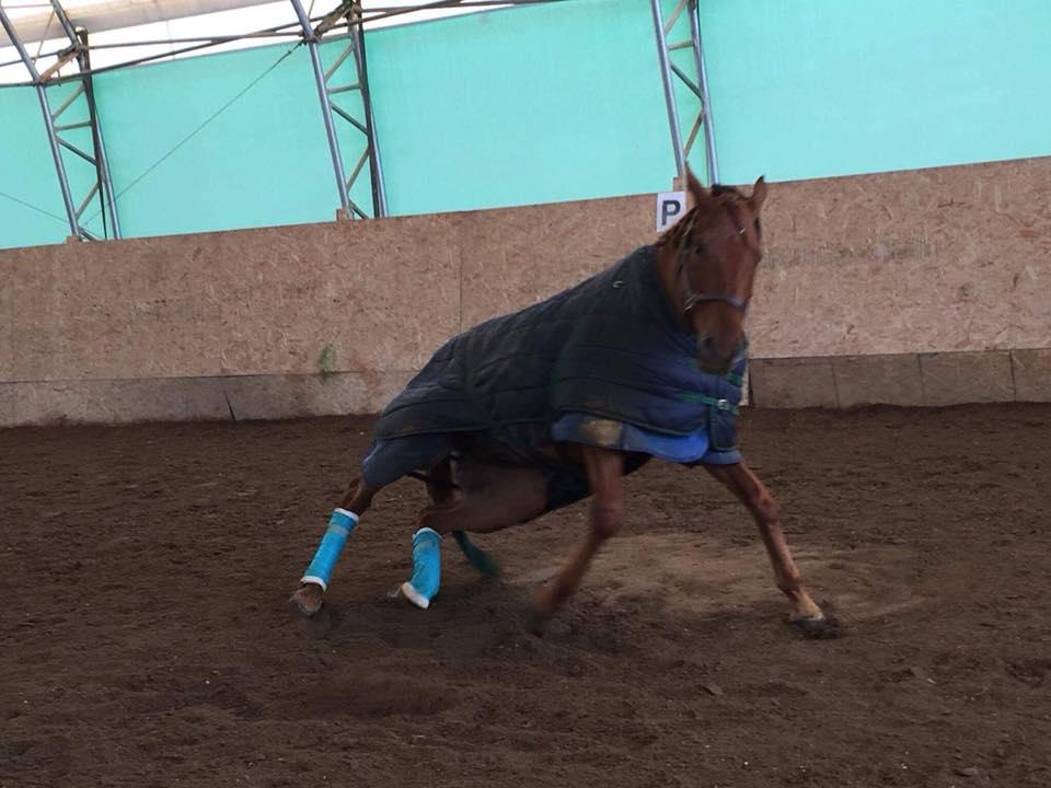
These malformed or compressed vertebrae press against the spinal cord and interfere with messages being sent by the brain. Interference with normal nerve transmission from the brain to the legs then leads to noticeable lack of coordination (ataxia). Ataxia is usually seen in the hindquarters. As the condition persists, weight loss and weakness may be noticeable. Clinical signs may appear suddenly or gradually, following known or suspected trauma, and may increase in severity until death, although death from wobbler syndrome is unusual. The horse may show periods of slight improvement but will never completely recover if aggressive treatment is not administered.
“Wobbler’s heel” is another clinical sign that occurs when the horse is in advanced stages of the syndrome and lack of coordination is evident. It is characterized by the horse reaching forward with the back foot and lacerating the bulbs of the heel of the front foot. This action can cause lameness, further complicating the syndrome.
Diagnosis
It is important to understand that multiple disease processes can lead to ataxia in a horse, including, in addition to wobbler syndrome, equine protozoal myeloencephalitis (EPM), West Nile virus (WNV), eastern and western equine encephalitis, equine herpes virus (EHV), rabies, trauma, equine motor neuron disease, and equine degenerative myeloencephalopathy (EDM). A thorough neurologic examination and diagnostic evaluation by a veterinarian in conjunction with owner observation and knowledge of the horse’s vaccination status are therefore crucial for the appropriate diagnosis.
Diagnosis of spinal cord damage requires quality radiographs of the cervical region. A radiograph measures the diameter of the cervical spinal canal in order to compare it against established ratios so that narrowed sites can be identified.
A myelogram is also used. It determines whether the compression is dynamic or static. This procedure is done under general anesthesia and involves injecting radio-opaque dye into the region behind the head in the area surrounding the spinal cord. Radiographs are taken
promptly after injection.
High-quality radiographs and a myelogram can be expensive and require special equipment and expertise. Horse owners and veterinarians can perform less costly tests to determine whether a horse has CVM.
The first sign that should prompt the owner to administer these tests is lack of coordination of the rear legs. Horses can compensate with their vision to coordinate the forelimbs, but they cannot visually compensate for their hind legs, so they will stumble.
Turning the horse in a small circle is a quick and easy test to detect wobbler syndrome. A horse suffering from the syndrome will swing the rear legs out while making the turn. The horse also will have trouble backing up. Instead of backing up their rear legs in a diagonal, two-beat fashion with their front legs, as is normal, the horse will back up with the front legs until it becomes awkward and then will hop backwards with the rear legs.
Another way of detecting wobbler syndrome is the sway test. Two people are necessary to administer it. One person walks the horse away while the other person grabs the tail and pulls the horse to one side. A normal horse will allow that to happen once before rapidly correcting itself. However, a horse exhibiting wobbler syndrome cannot tell where its limbs are, especially the hind limbs, thus you can easily pull the horse over to one side.
A third simple test used routinely is to pick up the horse’s tail. If absolutely no resistance occurs, the horse is having some type of problem with normal nerve function in its spinal cord. The weakness in tail resistance may be due to the pressure on the spinal cord caused by the malformation of the cervical vertebrae. Contact your veterinarian if you suspect your horse has developed wobbler syndrome or any other neurological disease.
Treatments
Horses with this syndrome can be treated with drug therapy, surgery, or aggressive management.
Certain drugs decrease the nerve tissue swelling and intracranial pressure. Some examples of these drugs include osmotic agents (for example, mannitol), dimethylsulfoxide (DMSO), and diuretics (for example, furosemide). Steroids are frequently used clinically for spinal cord and head trauma, but experiments have shown no benefit for wobbler’s syndrome because steroid therapy is targeted at treating the acute inflammation in the spinal cord that results from the compression but does not correct the underlying problem.
Surgery involves placement of a titanium basket within the vertebral bodies at the compression site, which serves to promote fusion of the vertebrae to relieve the compression. Th fusion of the bones aids in removing the instability caused by the malformation or malrticulations of the adjacent vertebrae. Surgery is an option in appropriate cases; it’s important to consider the number and location of compressions, degree of ataxia, duration of clinical signs, and the horse’s intended use. Surgery also requires a significant post-operative recovery time to allow for adequate healing. In the long term, complete recovery and return to normal athletic pursuits have occurred in 50-55% of cases Involving surgical treatment. Because of the later age of onset (2 to 4 years) of this disease, some animals may not necessarily returntoracing but may be able tocompete adequately in other fi lds.
Drug therapy and surgery are very costly and are not always practical for most horse owners. Aggressive nutritional management and controlled exercise coupled with early diagnosis have recently proven to be successful. The treatment is targeted at normalizing growth rates and allowing for proper bone remodeling within the cervical canal. Exercise is restricted in order to reduce the likelihood of trauma. Nutrition is focused on attention to appropriate calcium and phosphorus ratios and copper and zinc supplementation. A quality grass hay can serve as a source of roughage. This therapy takes time and patience before results are seen. Work closely with your veterinarian or equine extension specialist to establish your horse’s proper nutritional requirements.
Conclusion
Wobbler syndrome significantly impacts the athletic potential of affected horses and infl es the equine industry in a multitude of ways, both financial and emotional. Owners are often left with a difficult decision regarding treatment once a diagnosis is made. Euthanasia should be strongly considered, as wobbler syndrome can be unsafe for horse and rider.
Current molecular and genetic techniques are being used by researchers in efforts to answer the signifi ant question of the exact role of genetics in the disease’s development. Research also is under way on use of current imaging techniques to re-evaluate vertebral structure as well as bone metabolism.
All this research is focused on further elucidating the pathogenesis of wobbler syndrome in order to improve management and therapeutic practices of this disease.
