Neospora caninum Abortion in Cattle
Neospora caninum Abortion in Cattle
Neospora caninum is a protozoan parasite that is emerging as an important infectious cause of weak calves and abortion in cattle. N. caninum infections have been reported from most parts of the world with studies in the United States, New Zealand, the Netherlands, and Germany indicating that 12-45% of aborted fetuses from dairy cattle are infected with the organism. Exposure is common in US dairy herds ranging from 16% to 36% of dairy cows testing positive on serum. A distinguishing feature of the disease is abortion at 4-6 months of gestation, a unique time frame among infectious causes of bovine abortion. It is a very efficiently transmitted parasite with infection rates reaching 90% within some herds. Cows contract this parasite in one of two ways: 1) “Horizontally” by consuming feed or water contaminated with eggs (oocysts) from infected dogs and other canids or 2) “ vertical” transmission from cow to the fetus during pregnancy. An important feature of this parasite is that once it infects an adult cow or bull, a calf or a fetus, it is maintained as a life-long infection. Once infected, a cow can pass the organism through the placenta to her calf in every pregnancy throughout her lifetime. In some pregnancies, this fetal infection may result in abortion or weak calves. However the vast majority (95%) of calves born with the infection (“congenitally infected”) from positive dams are absolutely normal but remain infected for life. A heifer calf born with the infection can transmit the infection on to the next generation when she becomes pregnant, thus maintaining the infection within the herd. Vertical transmission (dam to calf) is known to be the major mode of transmission in cattle but both horizontal and vertical transmission are vital for parasite survival.
The way in which N. caninum causes abortion is complex and not fully understood. Abortion losses can occur after a primary infection (ingestion of eggs) but it is more commonly associated with reactivation of a persistent infection during pregnancy. Once the organism reaches the bloodstream of a pregnant cow (parasitemia), it invades the placenta and the fetus. Abortion is thought to occur by direct fetal and placental damage and/or the placental damage may cause release of maternal prostaglandins that cause luteolysis and abortion. Definitive diagnosis of abortion is through detection of the N. caninum organism in the fetal tissues, most consistently the fetal brain. There is no known drug to clear a cow of infection. A killed vaccine is available (NeoGuard, Intervet Inc) but studies suggest it has only a modest effect in reducing abortion risk and much uncertainty remains. Control is based on culling positive animals, preventing entry of infected replacements into the herd, and preventing likely routes of horizontal infection.
Life Cycle and Transmission of Neospora caninum:
Horizontal transmission of the organism occurs when a cow consumes an egg (oocyst) passed in the feces of a domestic dog, coyote, or other wild canid. Once inside the cow, the oocyst opens and releases tachyzoites that multiply rapidly and enter the bloodstream (parasitemia). Either the tachyzoites are transmitted through the placenta to the fetus or the immune system of the cow slows down the multiplication of the organism and they become encased in a shell (“encysted’). These “tissue cysts” are primarily found in the brain, spinal cord and muscles of cows but are thought to exist in the placenta as well. If dogs (or other wild canids) ingest these tissue cysts by eating a placenta, aborted fetus, or carcass of an infected cow, the organism can reproduce and form eggs (oocysts) in the intestines of dogs, beginning the cycle again. Deer are also known to form tissue cysts of N. caninum infective to dogs. Cow-to-cow transmission does not occur. Although not definitively proven, venereal transmission or by embryo transfer is considered highly unlikely.
Tissue cysts are primarily found in the central nervous system but can be in the muscle as well.
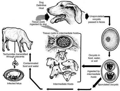
Illustration from Dubey JP: Neosporosis in cattle. Vet Clin North Am Food Anim Pract 21:473-483, 2005.
Vertical transmission occurs when the tissue cysts of persistently infected cows reactivate during pregnancy. It is believed that changes in the immune status of the dam allow tissue cysts to reactivate and release the organism to the bloodstream (parasitemia). In a majority of cases, vertical transmission leads to a healthy but congenitally infected calf. This contributes significantly to persistence within a herd by propagating the infection to successive generations.
Clinical Disease: Whether horizontally or vertically transmitted, bovine neosporosis is mainly a disease of the placenta and fetus. Adult cattle show no other clinical signs of disease. The fetus is most vulnerab
le to N. caninum before the 95th day of pregnancy and is unlikely to survive. In the middle third of pregnancy, the fetus may be able to mount an immune response that may or may not be sufficient to save it. In the third trimester, the fetus is usually clinically normal but infected for life. Abortion may occur as an epidemic or sporadic and occur at any time of the year. Unique characteristics of the disease include:
- A majority of abortions occur from 4-6 months gestation (range from 3 months to 8 months) with moderate rotting (autolysis) of the fetus. The fetus that dies in utero may also be resorbed, mummified or stillborn.
- The vast majority (95%) of calves born to positive dams will be born infected but normal.
- An infected calf may be born alive with neurologic signs, birth defects, and /or born weak and unable to stand.
- Cows can transmit the infection to their offspring in several consecutive pregnancies or intermittently.
Diagnosis: Examination of the fetus is necessary for a definitive diagnosis of abortion due to neosporosis. The best tissues to sample include brain, heart, liver, placenta, and also body fluids. Fetal brain is the most consistently affected organ and has the most characteristic lesion. Tests are available that can distinguish N. caninum from the two other protozoans that cause abortion in cattle, Toxoplasma gondii and Sarcocystis cruzi. A positive blood test from an aborting cow is only indicative of exposure to N. caninum and not diagnostic for abortion. There are several commercial cELISA blood tests that detect antibody to N. caninum that are rapid, inexpensive and consistent.
Control: N. caninum is efficiently transmitted vertically in cattle for several generations so culling of positive animals is one way to prevent this perpetuation of infection. In herds with a high prevalence of infection, blood testing the herd and selling the offspring of positive cows may be more economically feasible to reduce vertical transmission. Embryo transfer from positive cows to negative recipients can preserve valuable genetics safely. Testing of all purchased animals should be considered to prevent entry of positive animals in to the herd.
To prevent horizontal transmission, it is important to prevent exposure of the cows to feed and water contaminated with feces from dogs that may contain oocysts. Dogs and coyotes should not be allowed to eat aborted fetuses, fetal membranes, or dead calves. There is a killed parasite commercial vaccine (NeoGuard, Intervet) but there is no convincing data about the efficacy of this vaccine in preventing N. caninum abortion in cattle.
Neospora caninum is a protozoan that is becoming more widely recognized as an infectious cause of reproductive problems in cattle. Testing for this organism should be considered especially in herds that vaccinate routinely for infectious causes of abortion yet are still experiencing losses, particularly in the second trimester of pregnancy.
Author: Michelle Arnold, DVM
